Contact
Telephone:
(08158) 9220970
Fax:
(08158) 92209722
Address:
Bahnhofstraße 5
82327 Tutzing
Mail:
info@radiologische-praxis-tutzing.de
Surgery Hours
| Mon – Thu | 8:00 am to 6:00 pm |
| Fri | 8:00 am to 3:00 pm |
| As well as by arrangement | |
Services
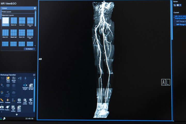
As a specialist practice for cross-sectional imaging examinations, we work with a 3Tesla high-performance MRI (Lumina, Siemens) and a 16-line spiral CT (Siemens), which can examine a slice thickness of 0.7 mm. We use high-resolution multi-channel coil systems in the MRI for the examinations of the individual joints with individually adapted examination protocols, depending on the problem. The findings are evaluated using workstations that automatically display the patient’s previous images to compare the findings. Even if you were a patient with us several years ago, you do not need to bring the previous images with you; thanks to a large image memory, all data is immediately available in digital form.
Magnetic resonance imaging (MRI)
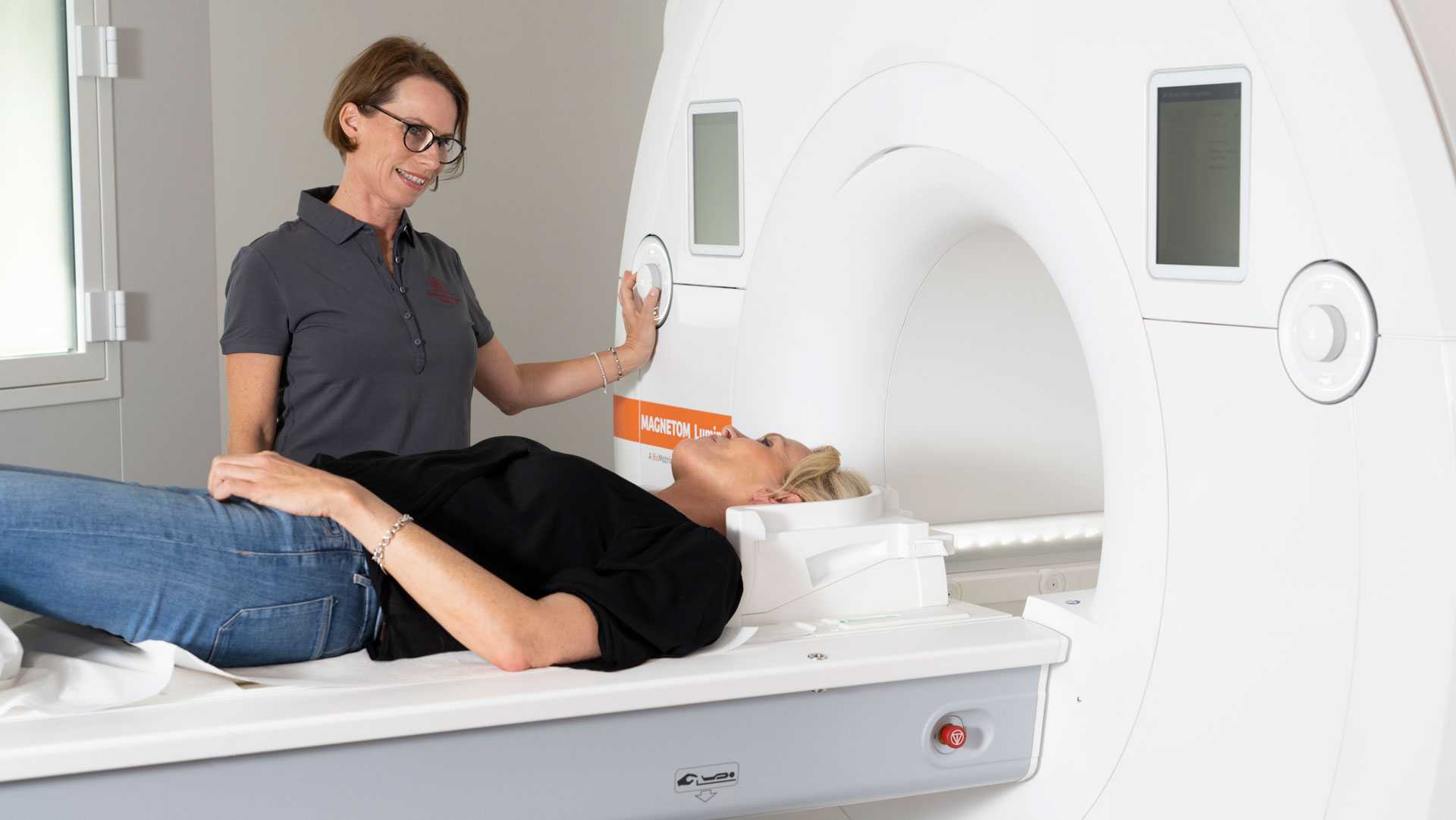
The SIEMENS Magnetom Lumina is a 3 Tesla nuclear magnetic resonance scanner of the latest design with a very large tunnel diameter (70 cm). This creates a new, pleasant feeling of space. The special lighting of the room and the MRI scanner makes patients feel comfortable and creates a positive examination atmosphere. Fear-free examinations are therefore the rule.
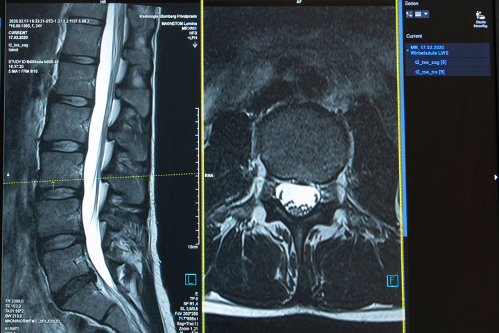
The most important prerequisite for recognising pathological findings is optimal image quality. This is the only way to diagnose even the smallest changes that cause discomfort (e.g. a small disc sequestrum that presses on the nerve) and thus to treat them correctly.
For this high-resolution image quality we use several components
- High-performance MRI (48-channel), which can depict even the smallest structures thanks to high field strength and multi-channel technology.
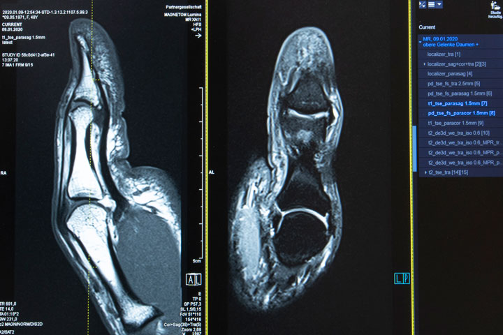
- The latest multi-channel coil systems, specially designed for the individual joints, which provide optimal image quality
- Special sequences (imaging protocols), individually adapted to the symptoms and the problem, which are characterised by a particularly thin layer thickness and a high level of detail recognition.
- Special workstations for the evaluation of findings, which can interactively process the findings even furth
Computed tomography
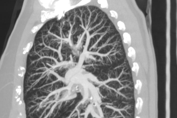
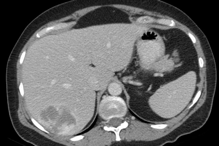
The 16-slice computed tomography (CT) scanner enables high-resolution imaging of bony structures of the spine and joints as well as temporally optimised imaging of internal organs and the skull with an optimal combination of low radiation exposure and high detail recognition. We use specially developed examination protocols for this purpose. A special concern for us radiologists is the reduction of radiation exposure; a CT examination is only performed after strict examination of the necessity.
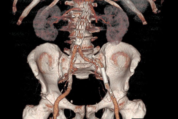
The volume data sets allow a 3-dimensional representation and with the help of the workstations and special software, virtual coloured 3D reconstructions are also possible. The correct position of metals after operations is also excellently possible without superimposition, despite the image distortions. In the case of complex bone fractures, the three-dimensional representation of the fracture fragments is a decisive help for the surgeon. Computed tomography is also used to assess delayed fracture healing and the stability of a healed fracture.
X-ray
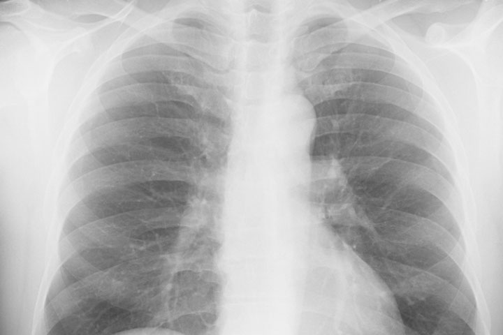
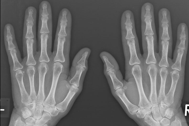
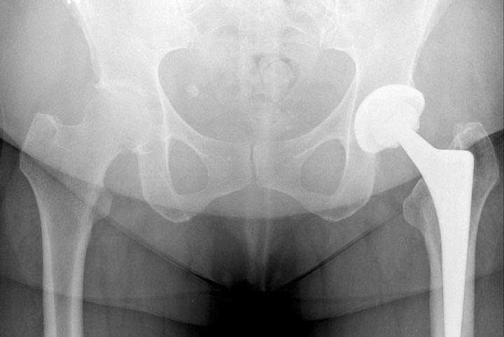
X-ray examinations are carried out digitally, the images are stored digitally like all other examinations, they can therefore no longer be lost, they can be sent electronically or via CD and are always available. The evaluation is done via workstations, so electronic measurements are possible. We would like to emphasise the possibility of carrying out digital spinal images, e.g. in the case of scoliosis, and loaded whole-leg images, e.g. in the case of pelvic obliquity or knee joint arthrosis with electronic measurement. For the preparation of an artificial joint replacement (hip, knee, shoulder), digital images with reference sphere for electronic size correction (MEDITEC company) can be made.
Contact
Telefon:
(08158) 9220970
Fax:
(08158) 92209722
Adresse:
Bahnhofstraße 5
82327 Tutzing
Mail:
info@radiologische-praxis-tutzing.de
Praxisöffnungszeiten:
Mo – Do 08:00 bis 18:00
Fr 08:00 bis 15:00
Sowie nach Vereinbarung
